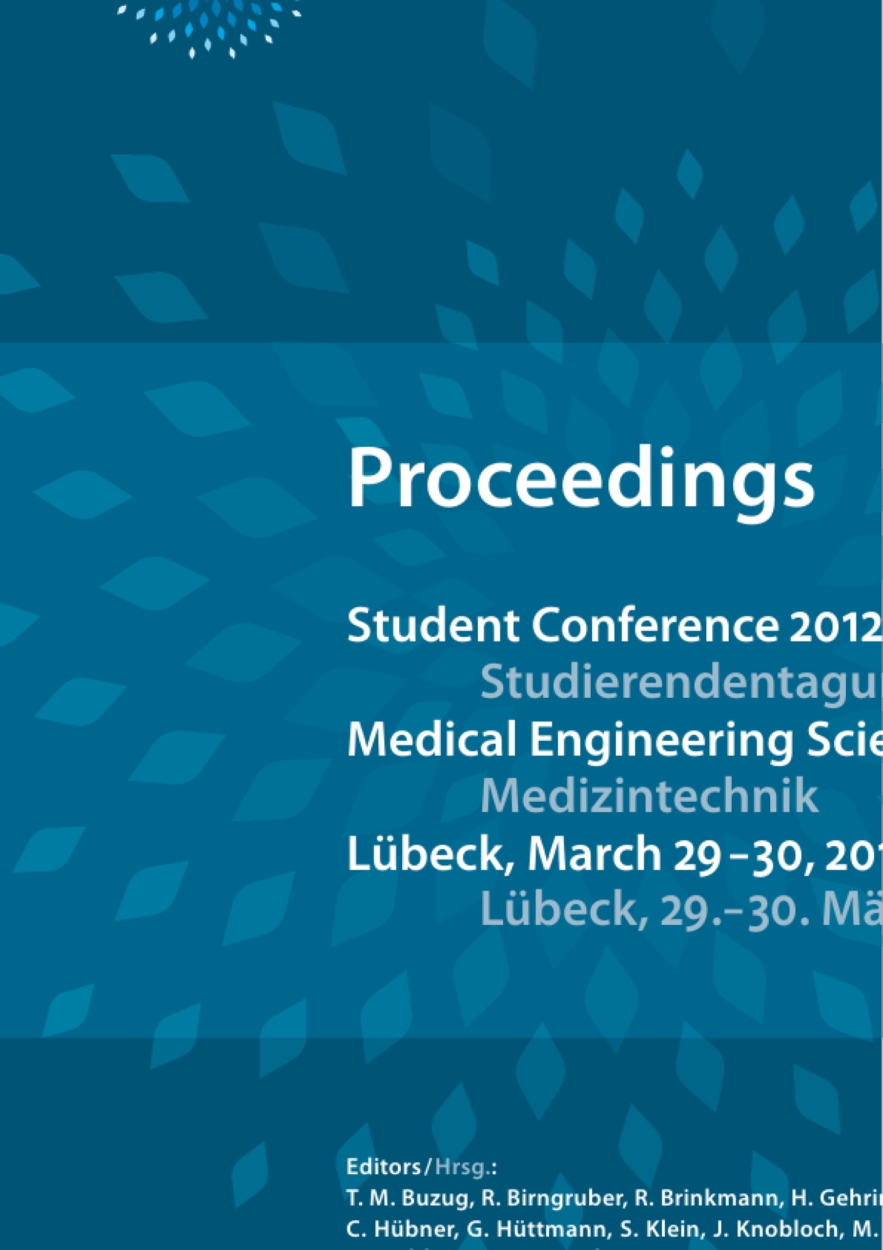
The Student Conference on Medical Engineering Science is an annual event at the BioMedTec Science Campus Luebeck. The Student Congress is organized by the University of Lübeck and supported by NORGENTA, the life science cluster agency in north Germany. Master students of programs related to medical engineering science present results of their recent research projects.
Die Studierendentagung Medizintechnik findet jährlich auf dem BioMedTec Wissenschaftscampus Lübeck statt. Der Kongress wird von der Universität zu Lübeck organisiert und von der norddeutschen Life-Science-Clusteragentur NORGENTA unterstützt. Studierende in Masterprogrammen der Medizintechnik und der Lebenswissenschaften präsentieren die Ergebnisse ihrer jüngsten Forschungsprojekte.
Inhaltsverzeichnis (Table of Contents)
- Preface and Acknowledgements
- Contents
- Biomedical Engineering I
- Development of a program to analyse and visualize ciliary beat frequency ex vivo
- Plug-in LED lighting for ureteroscopes
- CE certification of TTI Imaging
- EMG-based estimation of wrist kinematics using Fisher‘s linear discriminant analysis
- Material compatibility with different sterilization procedures
- Biomedical Engineering II
- Electrical Impedance Tomography Image Reconstruction with EIDORS
- Development and implementation of a method for producing directional solidified, electrospun hybrid structures as nerve guidance channels
- Spectral light modulation using a digital micromirror device (DMD) for the calibration of pulse oximetry sensors
- Multi-frequency Electrical Impedance Tomography for irreversible Electroporation
- Filtering cardiac artefacts from transdiaphragmal pressure for the validation of a non-invasive method to assess work of breathing
- X-Ray and Computed Tomography
- Emissivity factor comparison of different coatings for medical x-ray tube housings
- Models for osteoarthritis assessment from digital x-ray images of the lower extremity
- Phantom-based Determination of Noise Distribution in Computed Tomography
- Construction and Calibration of a Micro-CT Phantom for the Determination of Iron Oxide Concentrations in Ferrofluids
- Magnetic Particle Imaging
- Localization of small ferromagnetic samples in a magnetic particle imaging scanner
- 3-Dimensional FFP-MPI-Scanner Simulation using X-Space Theory
- Realistic Simulation of a Movable and Rotatable Field-Free Line in Magnetic Particle Imaging
- Magnetic Resonance Imaging
- Flexible Probe Positioning for Workbench Measurements on MRI Coils
- Curved saturation for spine imaging in magnetic resonance imaging
- Visualization of tumor tissue in the peripheral zone of the prostate using multi-parametric MR images
- Rotation estimation in k-space for different trajectories
- Biomedical Optics
- Development of a Novel Fractional Laser Device Utilizing a Tunable Cr2+:ZnSe Infrared Laser
- Full range Fourier domain optical coherence tomography via piezo-driven reference mirror
- Resection of human calcified aortic heart valves in vitro by using a Thulium laser
- Determining the accuracy and repeatability of a multidimensional eye tracker designed for laser refractive surgery
- Medical Image Computing
- Visualization of self-expanding stent systems and reject minimization
- Preprocessing of Spectral Retinal Images for Registration
- Application of Machine Learning Regression Techniques on Predicting Clinical Outcome in Primary Progressive Multiple Sclerosis
- Mathematical modelling of breast tumour growth and treatment
- Camera and tracking system calibration for image guided bronchoscopy
Zielsetzung und Themenschwerpunkte (Objectives and Key Themes)
The Student Conference on Medical Engineering Science 2012 aimed to provide a platform for master and diploma students to present their research findings in the field of medical engineering. The conference focused on a range of themes related to the field, including:
- Biomedical Engineering, including the development of new methods for analyzing and visualizing biological processes, and the creation of innovative devices for medical use.
- Imaging technologies, such as X-ray, computed tomography, magnetic resonance imaging and magnetic particle imaging, exploring new techniques for obtaining high-quality images and improving image quality through the use of innovative methods.
- Medical image computing, focusing on the development of new algorithms for analyzing and processing medical images, and the application of machine learning to predict clinical outcomes.
- Biomedical optics, investigating the use of lasers for tissue ablation and the development of new eye tracking systems.
Zusammenfassung der Kapitel (Chapter Summaries)
This preview excludes summaries of the conclusion or final chapter to avoid spoilers.
- Development of a program to analyse and visualize ciliary beat frequency ex vivo: This paper describes the development of a new program for analyzing ciliary beat frequency, which was implemented with MATLAB. The program improves the analysis of ciliary beat frequency of individual cells and generates a tool to visualize the changes in ciliary beat frequency of all cells present in the microscope field of view. The program was validated using transmission light microscopy on tracheae of mice ex vivo.
- Plug-in LED lighting for ureteroscopes: This paper examines the feasibility of using high-power LEDs as an alternative to xenon lamps in ureteroscopes. Experiments showed that LEDs can provide sufficient illuminance for small body cavities, and a first design of a mains-operated LED lighting device is depicted.
- CE certification of TTI Imaging: This paper describes the process of carrying out the CE certification for TTI Imaging software from EYETEC, a picture capture and processing software used in ophthalmology. The most important part of the certification is the risk analysis, which accounted for the bulk of the work. 40 risks from 30 different causes and 26 measures to minimize the risks were worked out in detail. After completing the conformity assessment procedure, EYETEC was allowed to sell TTI Imaging on the European market.
- EMG-based estimation of wrist kinematics using Fisher‘s linear discriminant analysis: This paper proposes an approach of using Fisher's LDA to estimate wrist kinematics, based on the assumption that linearly transformed electromyography (EMG) data could be used to estimate performed movements in free space. A mirrored-bilateral training strategy was used to investigate the clinical applicability of the algorithm for unilateral amputees. The results show the feasibility of using Fisher's LDA to estimate wrist kinematics.
- Material compatibility with different sterilization procedures: This paper examines the impact of different sterilization procedures on materials such as metals, plastics, elastomers and adhesives. The results show that the extent of changes is highly dependent on the sterilization procedure used. It was observed that adhesives are particularly sensitive to the effects of sterilization.
- Electrical Impedance Tomography Image Reconstruction with EIDORS: This paper describes methods and challenges in EIT to obtain measurement data and reconstruct spatial impedance distributions. Three different EIT example applications are presented: intracranial EIT, EIT for micro-vessel studies and EIT for irreversible electroporation (IRE) online feedback.
- Development and implementation of a method for producing directional solidified, electrospun hybrid structures as nerve guidance channels: This paper describes the development of a nerve guidance channel consisting of an inner chitosan scaffold and an outer fiber cover. The scaffold was produced with directional solidification to create a special pore structure, while the fiber cover was electrospun. The research focused on developing a special two-step method for electrospinning the scaffold. The results suggest that the developed nerve guidance channel could potentially aid in nerve regeneration.
- Spectral light modulation using a digital micromirror device (DMD) for the calibration of pulse oximetry sensors: This paper presents a novel calibration concept for optical sensors, such as pulse oximeters. The concept is based on the detection of the sensor’s optical signals, processing according to previously recorded measurements, and re-emission to the sensor to be calibrated. The core part of the concept is a light source with controllable spectrum, which was successfully achieved using a DMD.
- Multi-frequency Electrical Impedance Tomography for irreversible Electroporation: This paper presents a novel EIT system designed for the measurement, visualization and feedback of IRE. The developed system employs two electrode plates surrounding the target tissue for data acquisition and enables impedance spectroscopic measurements in real-time. The embedded system is in charge of data acquisition, preprocessing and transmission, while the host PC provides further filtering, data conditioning and the subsequent image reconstruction and display.
- Filtering cardiac artefacts from transdiaphragmal pressure for the validation of a non-invasive method to assess work of breathing: This paper deals with the effect of heartbeat artefacts on the measurement of transdiaphragmal pressure. Heartbeat artefacts were filtered off the data from three test subjects using a band rejection filter. The filtered and unfiltered data were analyzed and compared, resulting in an increase in the values for R² and chi_ok, which suggests that the filter is a useful tool for estimating heartbeat artefacts from the measured signal.
- Emissivity factor comparison of different coatings for medical x-ray tube housings: This paper investigates the emissivities of different coatings for medical x-ray tube housings. An experimental setup was developed to measure the temperature inside the housing samples, which were positioned vis-à-vis with an emitter of 1500◦C in ultrahigh vacuum. The results suggest that black chrome plating has the highest emissivity, while bright etching has the lowest.
- Models for osteoarthritis assessment from digital x-ray images of the lower extremity: This paper describes the development of models for quantitative OA assessment. The region of the intra-articular joint space of the hip, femorotibial and the upper ankle joint were modelled and implemented in a software, allowing for the calculation of quantitative parameters such as projected width and area of the joint cavity and force. The models were applied to eight segmented a.p. x-ray images of the lower extremity.
- Phantom-based Determination of Noise Distribution in Computed Tomography: This paper examines a simple idealized noise model for the measured intensity values in a clinical CT scanner. The results show that under real conditions and polychromatic radiation, even for low dose scans, the distribution of the intensity values corresponds to a Gaussian distribution.
- Construction and Calibration of a Micro-CT Phantom for the Determination of Iron Oxide Concentrations in Ferrofluids: This paper introduces micro computed tomography (micro-CT) as a new method for the determination of iron oxide concentrations of ferrofluids. A phantom and evaluation software were designed, which enable the user to determine the concentration without chemical altering the ferrofluid.
- Localization of small ferromagnetic samples in a magnetic particle imaging scanner: This paper studies a localization method for ferromagnetic samples using a magnetic particle imaging scanner. Phantom positions were reconstructed on a 1D-line in the center of the scanner system, obtaining a precision of around 1mm. The results show that the higher harmonics signal induced in the receive coils by the ferromagnetic samples contains sufficient spatial information for localization.
- 3-Dimensional FFP-MPI-Scanner Simulation using X-Space Theory: This paper describes the implementation of a three-dimensional MPI scanner simulation environment based on x-space theory. The simulation environment allows for FOV analysis, determination of resolution and phantom imaging. The results show feasibility by imaging two-dimensional and three-dimensional phantoms, confirming the theoretical resolution of the system.
- Realistic Simulation of a Movable and Rotatable Field-Free Line in Magnetic Particle Imaging: This paper presents a new MPI scanner design using a field-free line instead of a field-free point. The coil setup was simulated, and the field quality, heat generation and cooling system were optimized.
- Flexible Probe Positioning for Workbench Measurements on MRI Coils: This paper describes the integration of a robotic arm into an existing workbench setup for MRI coil measurements. A problem-oriented arm drive model was implemented, and the robotic arm was characterized in terms of reliability for a given configuration.
- Curved saturation for spine imaging in magnetic resonance imaging: This paper aims to design saturation pulses fitted to the spine geometry and the affected tissue in front of the spine. A two dimensional radio-frequency (RF) spatially selective excitation pulse was used to excite the specific saturation volume. The results show that the designed saturation pulse combined with an echo-planar imaging (EPI) sequence achieved a suppression of more than 97% in phantom measurements and 94% in subject measurements in brain.
- Visualization of tumor tissue in the peripheral zone of the prostate using multi-parametric MR images: This paper describes the development and evaluation of an automatic detection of tumor tissue in the peripheral zone of the prostate, based on different procedures of discriminating tumor from normal tissue. The results were evaluated on a cohort of 10 patients and demonstrate the potential of this method for easier localization and planning of treatment of prostate cancer using processed MR images.
- Rotation estimation in k-space for different trajectories: This paper investigates the influence of the data acquisition scheme on the quality of motion estimation in k-space. In particular, it investigates the feasibility of measuring rotation using the PCM from Cartesian k-space data and presents a stabilization method based on masking.
- Development of a Novel Fractional Laser Device Utilizing a Tunable Cr2+:ZnSe Infrared Laser: This paper describes the development of a novel fractional laser device using a tunable Cr2+:ZnSe infrared laser. The tunability of the laser allows for the investigation of wavelength-dependent laser-tissue effects and the development of customizable combinations of ablative and non-ablative FP treatments.
- Full range Fourier domain optical coherence tomography via piezo-driven reference mirror: This paper describes the development of a novel full range Fourier domain optical coherence tomography (FD-OCT) system using a piezo-driven reference mirror. The piezo-driven reference mirror allows for full range scanning, which potentially improves the accuracy and resolution of the OCT images.
- Resection of human calcified aortic heart valves in vitro by using a Thulium laser: This paper evaluates the cutting efficiency of an experimental high repetition rate pulsed Tm:YAG laser system for resection of calcified aortic valves. The results suggest that pulsed Tm:YAG radiation is promising for the resection of calcified aortic valves, while further investigation is needed for realizing minimally invasive resections.
- Determining the accuracy and repeatability of a multidimensional eye tracker designed for laser refractive surgery: This paper determines the accuracy and repeatability of a 6-dimensional eye tracker designed for laser refractive surgery. The results show that the eye tracker system achieves sufficient accuracy and repeatability for rotational and axial measurements to significantly reduce the effects of involuntary eye movements during laser refractive surgery.
- Visualization of self-expanding stent systems and reject minimization: This paper describes a system for visualization and quantization of catheters with self-expanding stents which are sorted out during the production process. The system aims to reduce the total reject by sensitizing employees to common failures and providing visualization in every production line.
- Preprocessing of Spectral Retinal Images for Registration: This paper presents a scheme for correcting spectral retinal images in terms of dust particles and uneven illumination. The information of a white reference image is used to perform the correction, and the results show that the presented method effectively prepares the spectral images for registration.
- Application of Machine Learning Regression Techniques on Predicting Clinical Outcome in Primary Progressive Multiple Sclerosis: This paper describes a machine-learning-based approach for the prediction of clinical features in a cohort of PPMS patients followed-up for 5 years. The results show that Gaussian Process (GP) can be used to predict clinical outcome with a high accuracy, especially for EDSS and TWT.
- Mathematical modelling of breast tumour growth and treatment: This paper presents a continuous mathematical model of early stage breast cancer progression and treatment response. The model considers a stepwise mutation pathway from a breast stem cell to a tumour cell and allows simulating the effect of different treatment strategies. The results suggest that TARGIT may be able to kill the remaining pre-malignant cells and prevent recurrence.
- Camera and tracking system calibration for image guided bronchoscopy: This paper presents a first approach to computing the geometric relationship between two different coordinate systems for image guided bronchoscopy. The goal is to find this relationship for two distinct C-arm positions, using CT images to mimic the real problem.
Schlüsselwörter (Keywords)
This preview includes key themes and concepts, such as medical engineering, imaging technologies, medical image computing, biomedical optics, and specific applications of these fields, such as the development of new methods for analyzing and visualizing biological processes, the creation of innovative devices for medical use, the improvement of image quality through the use of innovative methods, the prediction of clinical outcomes using machine learning, the use of lasers for tissue ablation, the development of new eye tracking systems, and the application of these techniques in various medical procedures.
Frequently Asked Questions
What is the Student Conference on Medical Engineering Science?
It is an annual event at the BioMedTec Science Campus Luebeck where Master students present their research in medical engineering and life sciences.
What are some key topics covered in the 2012 conference?
Topics included Biomedical Engineering, X-Ray and CT, Magnetic Particle Imaging (MPI), MRI, Biomedical Optics, and Medical Image Computing.
What is Magnetic Particle Imaging (MPI)?
MPI is an emerging medical imaging technique that uses magnetic fields to determine the location and concentration of superparamagnetic iron oxide nanoparticles.
How is machine learning used in medical engineering according to the abstracts?
Machine learning regression techniques were applied, for example, to predict clinical outcomes in patients with Primary Progressive Multiple Sclerosis.
What kind of laser research was presented?
Research included the use of Thulium lasers for resecting calcified heart valves and the development of fractional laser devices for medical therapy.
- Quote paper
- T. M. Buzug et al. (Author), 2012, Student Conference Medical Engineering Science 2012, Munich, GRIN Verlag, https://www.grin.com/document/200266


