Real-time Filtering of Physiological Tremor for Microsurgery. Physiological Tremor Robotic Compensation


Textbook, 2020

178 Pages, Grade: 2


Excerpt


Contents

List of Figures

List of Tables

1. Introduction
1.1 Context
1.2 Microsurgery and it’s Technical Challenges
1.3 Hand-held Robotic Instruments
1.4 Motivations
1.5 Contributions
1.6 Organization

2 Physiological Tremor Modeling: A Review
2.1 Physiological Tremor Genesis
2.2 Characteristics of Physiological Tremor
2.2.1 Time-domain Characteristics
2.2.2 Frequency domain Characteristics
2.3 Existing methods for Tremor Modeling
2.3.1 Amplitude-Domain Methods
2.3.2 Frequency-Domain Methods
2.4 Quantification of Physiological Tremor Modeling Methods
2.4.1 Physiological Tremor Database
2.4.2 Experimental Setup with Hand-held Instrument

3 Modeling of Physiological Tremor With Autoregressive (AR) Model
3.1 Introduction
3.2 Methodology
3.2.1 Autoregressive Modeling
3.2.2 Autoregressive Model with Least Mean Squares (LMS)
3.2.3 Autoregressive Model with Kalman Filter
3.2.4 Computational Complexity
3.3 Results
3.3.1 Optimal Initialization of AR model Hyper-parameters
3.3.2 Comparison Analysis
3.4 Experimental Evaluation
3.5 Discussion
3.6 Summary

4. Multi-step Prediction of Physiological Tremor
4.1 Introduction
4.2 Latency in Hand-held Instruments
4.3 Methodology
4.3.1 Multi-step prediction with BMFLC (MS-BMFLC)
4.3.2 Multi-step prediction with AR model (MS-AR)
4.3.3 Computational Complexity
4.4 Results
4.4.1 Optimal Initialization of Parameters
4.4.2 Comparison Analysis
4.5 Experimental Evaluation
4.6 Discussions
4.7 Summary

5 Machine Learning Techniques Based Multi-step Prediction of Physiological Tre
5.1 Introduction
5.2 Methodology
5.2.1 Conventional LS-SVM
5.2.2 Moving window LS-SVM (MWLS-SVM)
5.2.3 Tremor prediction with MWLS-SVM
5.3 Results
5.3.1 Parameter selection
5.3.2 Simulation results
5.3.3 Experimental validation
5.4 Discussions
5.5 Summary

6 Machine Learning Techniques based Characterization of Pathological Tremor For FES based Applicati
6.1 Introduction
6.2 Pathological Tremor Suppression with FES
6.3 Methodology
6.3.1 Proposed Approach For Pathological Tremor Filtering
6.3.2 Proposed Approach For Pathological Tremor Prediction
6.4 Pathological Tremor Database
6.5 Results
6.5.1 Optimal Initialization of mwLS-SVM Parameters
6.5.2 Pathological Tremor Filtering with mwLS-SVM
6.5.3 Pathological Tremor Prediction with mwLS-SVM
6.6 Summary

7. Conclusions and Future Directions
7.1 Conclusions
7.2 Future Directions

Referen

Summary

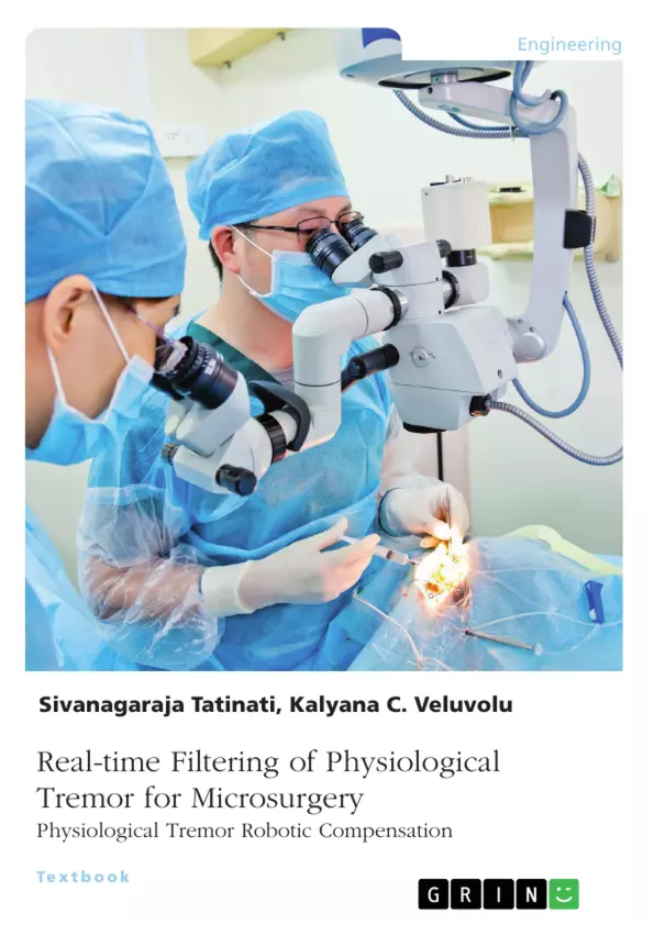
Precision, robustness, dexterity, and intelligence are the design indices for current generation surgical robotics. In microsurgeries, physiological tremor - an intrinsic hand motion with amplitude of 100pm - is a major impediment for surgeons' to perform delicate and fine motor tasks in sub-millimeter space. To augment the required precision and dexterity into normal microsurgical workflow by compensating the tremor in real-time, hand-held robotic instruments are developed. The working principle of a typical handheld instrument involves subsequent execution of three steps 1) sensing its own motion with inertial sensors, 2) filtering the involuntary motion from the sensed motion, and 3) actuate the surgical end-effectors (instrument tip) based on the filtered involuntary motions to compensate the tremor motion. Generally, digital filters are employed to filter out the noise components and subsequently extract the tremor motion from the whole motion. As a result, a time-varying and unknown delay in the range of 20 to 200ms (depends on the variant of the filter) is introduced into the tremor compensation proceedings, which in turn, adversely affects the tremor compensation performance. Ideally, zero phase lag between the actual tremor and extracted tremor motion is essential for hand­held instruments. This motivates development of new and innovative signal processing solutions, which can enhance the performance of hand-held instruments, in practice. We believe the key to achieve this goal is to go beyond the paradigm of conventional linear modelling techniques, which is limited to least squares solutions. This book proposes several solutions to overcome the existing issues and propose new solutions based on machine learning techniques for correction of phase delay.

List of Figures

Figure 1.1 Development of surgical robots for robotics-assisted microsurgeries: a) Tele­operated robotic surgical instrument - daVinci system (Intuitive surgical CA, USA) and b) cooperatively controlled robotic surgical system - steady hand (John Hopkins University)

Figure 1.2 Compensation procedure in a typical hand-held instrument

Figure 1.3 Thesis objectives

Figure 2.1 Multi-factorial origins of physiological tremor

Figure 2.2 Physiological tremor time series in three-dimensional space; normalized to zero mean and unit variance

Figure 2.3 Physiological tremor compensation with adaptive low pass filter: a) schemat­ic of adaptive low pass filter; and b) filtered tracking with adaptive low pass filter to illustrate the filter induced delay

Figure 2.4 Schematic of Fourier linear combiner

Figure 2.5 Schematic of weighted Fourier linear combiner

Figure 2.6 a) Schematic of band-limited multiple Fourier linear combiner; and b) frequency distribution of multiple FLCs

Figure 2.7 a) Experimental protocol: pointing task; and b) time-frequency characteris­tics of physiological tremor time series in Z-axis

Figure 2.8 a) Experimental protocol: tracing task; and b) time-frequency characteristics of physiological tremor time series in Z-axis

Figure 2.9 Schematic diagram for experimental validation of tremor prediction methods with hand-held instruments

Figure 3.1 Schematic of AR model with Adaptive Algorithms

Figure 3.2 Optimal order selection for AR model

Figure 3.3 Mean of estimation accuracy for various selections of R and Q for all trials and all subjects

Figure 3.4 Surgeon #4, Pointing task: filter coefficients (a) Tremor signal (b) AR- LMS coefficients (c) AR-KF coefficients; Solid line {wi,W2,W3} are for zero initialization, dotted line {w{,W2, Wg} are with selected initialization

Figure 3.5 Surgeon #4, pointing task (a) Estimation error with AR-LMS (b) Estimation error with AR-KF

Figure 3.6 Experimental setup with hand-held instrument to quantify the modeling of filtered tremulous motion with AR models

Figure 3.7 Surgeon #1, pointing task: real-time estimation with AR-KF (a) Estimation of Tremor; (b) Zoomed portion of estimated tremor (c) Filter coefficients (d) Tremor input and estimation error

Figure 3.8 Surgeon #1, pointing task: real-time estimation with AR-LMS (a) Es­ timation of Tremor; (b) Zoomed portion of estimated tremor (c) Filter coefficients (d) Tremor input and estimation error

Figure 3.9 Surgeon #1, pointing task: Prediction performance in presence of 8ms phase delay (a) AR-LMS (b) AR-KF

Figure 4.1 Latency in compensation procedure of a typical hand-held instrument

Figure 4.2 Frequency response of fifth order Butterworth bandpass filter (a) Phase response; (b) Input tremor signal and delayed tremor signal due to bandpass filtering

Figure 4.3 (a) Signal model (b) Schematic of proposed multi-step prediction approach

Figure 4.4 Multi-step prediction with BMFLC

Figure 4.5 Multi-step prediction with AR method

Figure 4.6 Performance analysis

Figure 4.7 Multi-step prediction for various prediction lengths: (a)BMFLC (b) AR (c) WFLC. Representation is standard deviation around mean

Figure 4.8 (a) Tremor signal (Subject #3, tracing task) ; (b) Predicton error with single-step prediction for BMFLC-KF; (c)-(e) Prediction error during the multi-step prediction with 20ms (10 samples) of prediction length for WFLC- KF, BMFLC-KF and AR-KF

Figure 4.9 Multi-step prediction accuracy for various sampling frequencies and various prediction lengths: (a) Prediction length 4ms (b) Prediction length 8ms (c) Prediction length 16ms d) Prediction length 20ms;

Figure 4. lCSchematic for experimental validation of developed multi-step prediction methods

Figure 4.1 Experimental results (a) Tremor signal (Subject #1, tracing task); (b) Prediction error with AR-KF; (c) Prediction error with MS-AR-LMS; (d) Prediction error with MS-AR-KF

Figure 5.1 Block diagram representation for tremor prediction with MWLSSVM

Figure 5.2 Latency in the tremor compensation

Figure 5.3 Offline training for MWLS-SVM

Figure 5.4 Flowchart representation of MWLS-SVM Ill

Figure 5.5 Grid analysis of %Accuracy for various values of C and cr2; N=500

Figure 5.6 Selection of signal history for offline training. Computational complexity (number of operations) of MWLS-SVM for every iteration (green in color)

Figure 5.7 Single-step prediction with MWLS-SVM

Figure 5.8 Performance of MWLS-SVM and LS-SVM for single-step prediction

Figure 5.9 Multi-step prediction with various induced delays for all methods

Figure 5.1 QMulti-step prediction performance in the presence of various induced delays

Figure 5.1 IScatter plots

Figure 5.12 Multi-step prediction in the presence of unknown delay

Figure 5.13 (a) Surgeon #1 (pointing task) (b) Prediction error due to unknown delay (c) Prediction error with MS-BMFLC-KF (d) Prediction error with LS-SVM (e) Prediction error with MS-AR-KF (f) Prediction error with MWLS-SVM

Figure 5.14 Performance analysis of all methods in the presence of unknown delay

Figure 5.1 Experimental procedure

Figure 5.1 (Prediction performance in presence of unknown delay a) tracing task of Surgeon #1 and b) tracing task of Novice Subject #1

Figure 6.1 Delay in tremor suppression procedure based on FES

Figure 6.2 Block diagram representation of pathological tremor filtering and prediction

Figure 6.3 Multistep prediction of pathological tremor with mwLS-SVM

Figure 6.4 Multistep prediction with mwLS-SVM (a) Whole motion (Subject #1, action tremor); (b) Tremulous motion obtained after bandpass filtering ; (c) Prediction error obtained due to 80 ms delay; and d) multistep prediction error obtained with mwLS-SVM

Figure 6.5 Pathological tremor filtering and prediction with mwLS-SVM (a) Whole motion (Subject #1, action tremor); (b) Filtered voluntary motion with mwLS-SVM (c) Extracted tremulous motion together with mwLS-SVM based multi-step predicted tremulous motion ; and (d) Prediction error obtained for prediction horizon 60 ms

List of Tables

Table 3.1 Adaptation Schemes

Table 3.2 Computational complexity

Table 3.3 Methods & Parameters

Table 3.4 Settled filter coefficients (M ± SD) for 5 surgeons (S) and 5 novice subjects (NS) with AR-KF

Table 3.5 Estimation performance (M ± SD) for 5 surgeons (S) and 5 novice subjects (NS)

Table 3.6 Comparison with existing methods

Table 4.1 Performance of BMFLC-KF 1 in presence of phase delay

Table 4.2 Adaptation Schemes

Table 4.3 Computational complexity

Table 4.4 Methods & Parameters

Table 5.1 Optimal ranges for C and cr2 selection with various values of N

Table 5.2 Methods & Parameters

Table 5.3 Comparison analysis with existing single-step prediction methods

Chapter 1

Introduction

1.1 Context

Anthropomorphic design constrains humans to accomplish micro-manipulation tasks that demand dexterity and precise spatial resolution in upper limb maneuvers. The primary factor that limits the precise spatial resolution of maneuvers in humans is small-magnitude rhythmic involuntary movements. These movements are concomitant with all sorts of voluntary muscle contractions, and are named as Physiological tremor [2, 3, 4, 5], Physiological tremor has been extensively investigated over the last five decades and is attributed as a ubiquitous property of humans [2, 5]. It is believed that physiological tremor originates from both mechanical and neuromuscular sources [3, 4]. However, a lucid view of its origin is still lacking. The amplitude of physiological tremor is usually in millimeter range and barely visible to the naked eye [2, 3, 4, 5]. In frequency domain, the dominant peak of physiological tremor lies in the range of 6 to 12 Hz [3, 4, 6, 7]. By virtue of its biological origins, physiological tremor is non-stationary in nature 5. Experimental studies conducted on physiological tremor have determined that it is a linear stochastic process [5, 8]. Further, it’s modulation with voluntary motion in time-domain is additive, the magnitude of tremulous motion is superimposed on the magnitude of voluntary motion [5, 8, 6].

The effect of small-magnitude physiological tremor on normal daily living activities is benign. However, for the activities where the magnitude of voluntary motion is same as that of involuntary motion and for the activities that deal with a workspace in millimeter range, the effect of physiological tremor is adverse 9 . For example, in the arena of microsurgery where surgeons perform complex maneuvers in sub-millimeter- sized anatomy, physiological tremor has deleterious effects [9, 10].

1.2 Microsurgery and it’s Technical Challenges

Surgical procedures under a microscope was initially proposed in 1962, to aid the repair of blood vessels (vascular surgery) 11. During that era, the limitations of these procedures were attributed to the capabilities of imaging techniques rather than the surgeons’ dexterity. In recent decades, technological developments in imaging techniques have improved the quality of surgical procedures and pushed the limits of surgeon’s dexterity and precision [9, 5, 12]. Further, the lack of required spa­tial resolution in surgeon’s maneuvers limits the number of feasible micro-surgical procedures.

Opthalmologic surgeries are very complex in nature because of the eye’s delicate structure and rich network of tiny and sensitive blood vessels. Success in these procedures depends on an efficient repair of damaged vessels/tissues without damaging the surrounding vessels/tissues [9, 10]. For instance, vitreoretinal microsurgery deals with removal of approximately 20 fim thick membranes from retina [10, 12]. While performing a vitreoretinal microsurgery, oscillaitons of magnitude ~ 50 [im peak-to- peak in each principal axes were measured at instrument tip [12, 13, 14]. According to 15, required precision for the surgical instrument tip in vitreoretinal micro-surgical procedure is approximately 10 /d/m. Other micro-surgical procedures such as removing tumors from neurons (neurological surgery), vascular repair (micro-vascular surgery), and treating retinal vein occlusions etc, demand dexterity and precision in surgeon’s maneuvers up to sub-millimeter range.

The spatial resolution required to perform successful micro-surgical procedures limits the number of qualified microsurgerons. Robots, on other hand, are famous for repeating maneuvers with high spatial resolution (precision) and dexterity [16, 17, 18]. The attributes possessed by robots are invaluable in the arena of microsurgery, es­pecially when dealing with sub-millimeter anatomies 18. In an attempt to perform successful micro-surgical procedures, robotics-assisted surgical procedures were de­veloped [19, 20]. The main objective of the robotics-assisted surgical procedures is to augment the respective advantages of robots such as repeating maneuvers with high precision and dexterity etc with the respective advantages of surgeons such as high level cognitive power etc. Thus, in last few decades, robotics-assisted surgery has become a dynamically growing research area with worldwide clinical uptake 21.

The first ever usage of robot in microsurgery (neuronal) was reported in 1985 20. An autonomous robot was employed to hold the biopsy cannulae. Over the last three decades, robotics-assisted micro-surgical procedures have evolved from autonomous to tele-operated and then to co-operative robots [21, 22], as shown in Figure. 1.1. In tele-operated robotics-assisted surgeries, the tremulous surgeon hand was replaced by a robotic arm. Surgeon controls the patient-side slave robotic arm remotely through manipulators at master-side console, for example the daVinci system (Intuitive surgical CA, USA). In co-operative controlled robots assisted surgeries, both the robot and surgeon hold the instrument together and perform manipulations. This co-operation enables surgeons to have real-time control over the instrument with limited degrees of freedom and improves the precision of instrument positioning, for example Steady-hand robot (John Hopkins University).

Tele-operating robotics-assisted surgeries were attributed with clinical success. Nonetheless, the hospital uptake of these instruments was not encouraging because of their bulky size, high cost, long preparation time for surgery and special training required to utilize these instruments. Further, surgeons prefer performing surgeries with instruments that can offer natural and familiar feel of conventional surgical instruments. Considering these limitations, in recent years, the research on surgical robots has shifted towards developing smart surgical-robotic instruments that are less obstructive, similar in physical specifications to conventional surgical instruments, and intelligent enough to compensate the involuntary motions. This kind of surgical robots are named as hand-held robots. This dissertation focuses on hand-held robotic instruments applicable for microsurgical procedures.

1.3 Hand-held Robotic Instruments

The first ever hand-held robotic instrument for the compensation of physiological tremor was developed in the year 2000 and was named as Micron 23. The main advantages of hand-held robotic instruments when compared to the other variants of surgical robots are: compact in size, easy to perform maneuvers in free-space similar to the conventional surgical instruments, and cost effective 22. Furthermore, hand-held instruments can mitigate the circumstances that lead to catastrophic failures during surgical procedures, because even in case of failure the hand-held instrument is not capable of displacing its tip more than a few hundred micrometers. If compensation of hand-held instrument is shut down, it simply becomes a passive hand-held instrument such as the surgeon has worked with for years. With these advantages, physiological tremor compensation with hand-held instruments have became a rapidly growing research area.

Physiological motion compensation mechanism with a typical hand-held instru­ment is shown in Figure 1.2. It is engineered with three distinct units: sensing unit, filtering and modeling unit, and compensation unit. The sensing unit houses inertial sensors such as accelerometers to sense the maneuvers made by surgeon during the surgical procedure. The sensed motion comprises of both the surgeon’s intended motion and concomitant involuntary motions. First, the filtering and modeling unit converts the sensed motion in acceleration domain to position domain by performing double numerical integration. This unit further employs frequency selective linear filters to separate the involuntary motion components from the sensed motion. Finally, adaptive methods such as band-limited multiple Fourier linear combiner (BMFLC) are employed to model the filtered physiological tremor components. The compensation unit then utilizes the modeled tremulous motion components to generate a control signal for the piezo-electric actuators that are housed at the tip of the instrument. Based on the generated control signal, the actuators manipulate the surgical tool tip position and hence compensates the involuntary motion.

Over the years, several variants of hand-held instruments have been developed for increasing the compensation capabilities of the instrument. Considering precision, weight and dimensions as constraints, inertial sensors/lasers and piezo electric actua­tors/polymeric metallic components based actuators are employed in the sensing and compensation units respectively. The variants of hand-held instruments are Micron 23, iTrem 24, visual-aided Micron 25, iTrem2 26 and AID 27. During this evolution, the sensing and compensation units became more accurate and robust. Further, the weight of the instrument has significantly scaled down. The latest variants iTrem2/Micron weighs comparable to the conventional surgical instrument [24, 25].

Despite the advantages, there are several technical challenges in the development of hand-held instruments for accomplishing accurate compensation of physiological tremor in real-time [12, 24, 25]. A significant challenge is accurate and real-time separation of surgeon’s intended motion components and involuntary motion components from the sensed motion.

1.4 Motivations

The three units of hand-held instruments execute in sequential manner to compensate the physiological tremor, as shown in Figure. 1.3. First, the sensing unit senses the whole motion in acceleration domain. Then the filtering and modeling unit converts the sensed motion into position domain and then filters out the tremulous motion components. The filtered tremulous motion components are then modeled and predict­ed using adaptive methods to generate the control signal for the compensation unit. Finally, according to the control signal generated the compensation unit manipulates the tip position.

Over the past decade, several signal processing methods have been developed and customized for hand-held instruments to model and predict the filtered tremulous motions [12, 28, 29, 30]. Of all the methods, weighted Fourier linear combiner (WFLC) and BMFLC are empirically proved to be accurate prediction methods [28, 29]. WFLC updates both the amplitude and frequency components of the model iteratively. On the other hand, BMFLC limits the signal’s bandwidth of interest and updates the amplitude of the frequency components in that bandwidth iteratively. In [28, 29] it was concluded that prediction performance of both methods relies on the optimal initialization of their parameters. To do so, both methods require prior knowledge on subjects tremor characteristics. If the prior knowledge is not available, then the whole frequency band of involuntary motions i.e., 2 to 20 Hz has to be considered. In this scenario, modeling is susceptible to unwanted components and hence prediction is not accurate.

The prediction performance of WFLC and BMFLC methods on filtered tremulous motion alone, with optimally initialized parameters, was reported as approximately 82% and 90% respectively [12, 24, 29]. However, the overall compensation accuracy with BMFLC dropped to 39% upon experimental validation 29. In 1, the decrease in compensation accuracy was attributed to the delay induced by linear filters (band-pass filter) employed to separate voluntary motions and involuntary motion components in the filtering and modeling unit. In [1, 31] it was further stated that, accurate compensation of physiological tremor components could be achieved only if the separated physiological tremor components were accurate and in-phase with the sensed motion. If the filtered components are not in-phase with the sensed motion, the manipulations provided by the compensation unit might amplify the physiological tremor components instead of compensating them. This implies that assigned tasks for all three units in hand-held instruments have to be accomplished in one sampling unit without any phase delay.

The hand-held instruments require linear filters to filter out the notorious integra­tion drift obtained while converting the sensed motion from acceleration domain to position domain, besides filtering the tremulous motion components [24, 28]. Further, these filters are required to filter out the other involuntary motions such as chorea, jerk etc [24, 28]. As such, employing linear filters serve dual purpose and it is mandatory.

Linear filters in general need taps (future values) to filter out the components in the desired bandwidth 32. As a result of causality, the filtered tremulous motion components are delayed compared to the sensed motion components.

According to 33, delay as small as 30 ms degrades the overall performance in human-machine control applications. To add to the technical difficulties, tremulous motion is non-stationary in nature and has high frequency 6 to 14 Hz. Thus, a delay as small as 10 ms brings phase shift of 45° and it will adversely affects the compensation capabilities of hand-held instruments. Consequently, a zero-phase adaptive filter suitable for this application has to be both adaptive and predictive. Since the intended application is microsurgery, the filter has to be robust to unwanted noise, further, the compensation is performed in displacement domain and therefore the algorithm should provide drift free position information for compensation of tremor.

Motivated by these challenges, the work conducted in this dissertation focuses on issues that are related to the accurate and real-time separation of physiological tremor components from the sensed motion. The main objectives of this dissertation are (shown in Figure 1.3):

i. developing tremor modeling methods that do not require any prior knowledge of subjects tremor characteristics,
ii. developing new algorithms to accurately model and predict the separated physio­ logical tremor in the presence of delay, and
iii. developing new procedures to separate the physiological tremor from the sensed motion accurately in real-time.

1.5 Contributions

This dissertation mainly focused on developing new algorithms and techniques for accurate modeling and prediction of physiological tremor components (the filtering and modeling system) for the hand-held instruments. The methods developed in the course of this dissertation were validated with the physiological tremor database collected from micro-surgeons and novice subjects. Further, the methods were experimentally validated with the bench tests conducted on hand-held instrument, iTrem.

i. To develop a simple and robust method that can model tremulous motion without any prior knowledge, we explored the application of autoregressive (AR) models for modeling tremulous motion. To adaptively update the AR coefficients, we employed adaptive filters such as least-mean square (LMS), and Kalman filter (KF). Though estimation with AR models was accurate, AR coefficients required ample amount of time to settle. This initialization period affected the tremor estimation accuracy. Thus, we conducted a study on the physiological tremor database and identified optimal initialization for AR coefficients. The initialization showed a significant improvement in estimation accuracy. Tremor modeling with AR methods was experimentally validated on iTrem.
ii. Analysis was conducted on the employed linear filters and the compensation procedure of hand-held instruments, identified that the filtered tremulous motion with a linear filter delayed by 12 to 20 ms when compared to the actual tremulous motion. Despite accurate modeling of tremulous motion with AR methods or BMFLC, control signal generated with delayed physiological tremor components amplified the involuntary components at the instrument tip instead of compen­sating them. To counter the limitations with phase delay, multi-step prediction of physiological tremor was proposed. Multi-step prediction of physiological tremor was challenging because of its stochastic nature and high frequency range. To perform multi-step prediction of physiological tremor, AR model and Band-limited multiple linear Fourier combiner (BMFLC) were customized. Developed multi-step prediction approaches were experimentally validated.
iii. The developed multi-step prediction methods significantly improved the estimation accuracy of tremulous motion in the presence of delay. However, these methods assumed physiological tremor as a stationary signal in the given prediction horizon and required a prior knowledge on prediction horizon. In contrast, physiological tremor is non-stationary in nature and the delay obtained after filtering stage depends on both subject and time. To address these issues, we developed a horizon- free prediction method with least squares support vector machines (LS-SVM). Since, LS-SVM prediction performance is susceptible to non-stationary signals, we developed a moving window based online training scheme for LS-SVM, named as mwLS-SVM. The mwLS-SVM updates the nonlinear mapping sequentially with every available new sample and hence adapts to the time-varying characteristics of tremor.
iv. To enhance the pathological tremor suppression capabilities with functional electrical stimulation (FES), the above developed methods and procedures were employed for active filtering and prediction of pathological tremor.

While the targeted application of this dissertation is in microsurgery, the principle of the approach and the theory of operation is universal and can be extended for other micro-manipulation tasks like cell manipulation in bio-tech industry and gun-sights or hand-held military equipment etc.

1.6 Organization

The remaining chapters in the dissertation are organized as follows:

In Chapter #2, a brief description about physiological tremor, its genesis and a literature review on methods developed for physiological tremor modeling in hand-held instruments is provided. Further, a brief description about the physiological tremor database and the experimental setup employed to validate the methods developed in the later chapters of this dissertation is provided.

Chapters #3, # 4, #5 and #6 form the core part of this dissertation. In Chapter # 3, modeling of physiological tremor with autoregressive models and its experimental validation of AR methods with iTrem are described. In Chapter t^4, the analysis conducted to identify the phase delay in the compensation procedure is detailed. It is followed by the developed multi-step prediction approaches and its experimental validation. In Chapter #5, the necessity of developing a horizon-free multi-step prediction method and its experimental validation are described. In Chapter y^6, modeling and prediction of pathological tremor with the above developed methods for functional electrical stimulation based suppression are described.

Finally, Chapter #7 concludes the dissertation by reviewing the accomplishments and limitations of this research, and gives an account for future work.

Chapter 2

Physiological Tremor Modeling: A Review

The purpose of this chapter is to provide a comprehensive review and background knowledge on physiological tremor genesis. In this chapter we further discuss about time-domain and frequency-domain characteristics of physiological tremor. It is followed by a literature review on recent works that has contributed towards accurate modeling of physiological tremor for hand-held instruments.

2.1 Physiological Tremor Genesis

The voluntary contraction of any skeletal muscle is accomplished by activation of a functional unit called motor unit. The motor unit comprises of motor neuron and the muscle fibers which it innervates. The discharge of a motor neuron creates a twitch in the innervated muscle fiber and simulates the contraction 34. The size of the motor unit and the number of muscle fibers that it innervates contribute to the force of muscle contraction. Activation of motor units is usually asynchronous, hence the contraction of muscle is not smooth. These asynchronous activations cause involuntary, rhythmic, and stereotyped movements which are superimposed on the voluntary contraction of muscle 35. These involuntary movements are named as physiological tremor.

The amplitude of physiological tremor is usually very small and hardly visible to naked eye. The frequency components of physiological tremor depends on the muscle group associated with limb segment. The frequency components are usually in the range of 2 to 30 Hz [36, 34, 37]. Researchers have attributed physiological tremor as a ubiquitous characteristics of voluntary muscle contraction 38. The first ever proof of superimposed tremulous twitch existing in a voluntary muscle contraction was reported in 1886 39. Ever since the electro-physical recordings of tremulous motions were acquired, study on the genesis of physiological tremor has been extensive. Technological developments in recent times have aided the study on physiological tremor to a great extent [40, 41].

Experimental and neurological studies conducted on a large volume of normal subjects revealed that physiological tremor is a combined outcome of four distinct sources (oscillations) and the contribution of each source to physiological tremor depends on either limb segment is at rest, held against gravity, or in motion 3. The small amplitude tremor that exists when limb segment is at rest is considered as rest tremor. The source for rest tremor is identified as ballistocardiac impulses 42. Postural tremor exists when limb is out-stretched or hold in a position against gravity. The origins of postural tremor is attributed to both mechanical oscillations and stretch reflex loop [43, 44, 45]. The presence of tremor while performing actions or goal-oriented tasks is named as motion tremor. The motion tremor has multi­factorial origins central oscillations (central nervous system) and mechanical oscillations (variations in mechanical properties) [46, 47].

The multi-factorial origins that could account for physiological tremor are depicted in Figure. 2.1. A brief description about the mentioned origins (oscillations) is defined as the following:

Mechanical Oscillations

The intrinsic resonant frequency of any limb segment depends on its stiffness (K) and moment of inertia (J). The resonant frequency (/o) can be given as 45:

Abbildung in dieser Leseprobe nicht enthalten

The resonant frequency of each limb segment is different, for example the resonant frequency of metacarpal-phalangel joint (unloaded) is in the range of 12 to 30 Hz, while for wrist and elbow it is 8 to 12 Hz and 3 to 5 Hz respectively [5, 45]. The variations in resonant frequency of any limb segment relies on the physical properties of bone, soft tissue, and muscle group associated with that segment 6. According to (2.1), the resonant frequency of any limb segment will change upon adding external weight or applying force. In [48, 49], it was stated that, adding external weight will increase the inertia of the segment, consequently decrease its the resonant frequency. Experimental studies determined that for most of the subjects the frequency components due to the mechanical oscillations are the dominant components in their physiological tremor spectrum 6.

Balistocardiac impulses

The blood pumping mechanism of heart in particular the thrust while contraction is considered as another source for physiological tremor 50. It was believed that the blood circulation mechanism exerts significant force on all body parts and this force causes perturbation on resonant frequency of body parts that could account for physiological tremor [50, 51]. The frequency components of balistocardiac impulses are the dominant components in the physiological tremor spectrum if the limb is at rest 50.

Stretch-Reflex Oscillations

Central nervous system (CNS) controls muscle actions through reflex loops. There are peripheral loops which connect muscles to spinal cord with a feedback, and central loops connecting muscles to higher segments of spinal cord, the brain, and the brain stem [42, 44, 45]. Depending on the reflex gain and the transmission delays of the loops, oscillations that account for physiological tremor prevail [44, 34]. For example, the simplest loop is from the muscle spindle to the muscle fiber through motor neurons and motor axon 52. The time required to complete the peripheral loop (spinal segmental stretch) in the finger i.e., the time between EMG onset and detectable movement at afferent receptors is 50 ms [52, 42]. This transmission time creates oscillations at the frequency 10Hz.

The studies conducted on large volume of subjects revealed that the contribution of stretch-reflex oscillations to physiological tremor is very small. These oscillations however modulate the amplitude of physiological tremor with an unknown index [52, 42].

Central Oscillators (Central Nervous System)

The first ever hypothesis on the genesis of physiological tremor was attribute to central oscillations (oscillations due to CNS) [39, 3, 2], The contribution of central oscillations either directly or indirectly has been proved by studies on normal subjects [39, 44, 53]. For example, the studies conducted on deafferented patients (with no sensory feedback loops) have revealed the existence of postural physiological tremor [54, 55] and the experiments conducted on normal subjects involving complicated finger movements have revealed no effect on tremor components upon altering mechanical properties of finger [43, 46].

Experimental studies have confirmed the existence of 8 to 12 Hz frequency components in muscles groups such as biceps, extensors, and intrinsic hand muscles with widely varying mechanical properties [46, 47]. These frequency components are preserved independently on loading effect 44. It is further suggested that the central oscillations are the primary reason for frequency-variations of physiological tremor [44, 45, 56, 57].

2.2 Characteristics of Physiological Tremor

The two most common characteristics of physiological tremor have been its amplitude and frequency. These two measures however are not capable of clearly characterizing the nature of physiological tremor. Thus, researchers have proposed other charac- teristics/measures that could be more discriminative and explain the dynamics of physiological tremor. The measures are usually evaluated from the kinematics or force signals. These measures can be broadly divided into time-domain and frequency­domain characteristics [58, 59, 60, 61]. A detailed description about the calculation of these measures can be found in [60, 61]. In these works, the tremulous motion was acquired as a time series and then normalized to zero mean and unit variance, as shown in Figure. 2.2.

2.2.1 Time-domain Characteristics

The characteristics evaluated in time-domain to determine the nature of physiological tremor time series are linearity, stochasticity, and distribution of the data points.

Linearity

In general, linearity tests on time series are based on the assumption that the dynamics of a process can be driven by Gaussian noise. These tests analyze the time series structure by its third moments. In the field of time series, test of Keenan 62 and test of Tsay 63 have been established to determine the linearity. The analysis conducted on tremor time series in [64, 8] determined that the dynamics of physiological tremor were linear and not chaotic.

Stochasticity

The stochastic nature of physiological tremor time series was evaluated by computing the correlation dimensions (embedded dimension) 8. According to Taken’s theorem, if the correlation dimensions have few degrees of freedom then the time series is deterministic. In case of many degrees of freedom (infinite), then the time series is stochastic in nature. In 8, the correlation dimensions for physiological tremor were calculated and concluded that the dynamics of physiological tremor were stochastic in nature.

Features of the distribution

The analysis conducted on distribution of physiological tremor time series determined the Gaussianity in tremor dynamics [64, 8]. In 64, the third moment (skew) and fourth moment (Kurtosis) are computed for the physiological tremor distribution. The cumulative distribution obtained for the third moment of tremor time series showed zero skewness with slight deviation 64, The measure fourth moment (kurtosis) for tremor time series showed a good agreement with Gaussian distribution (0.798) 65.

The dynamics of the physiological tremor time series were therefore determined as linear Gaussian process. This deduction is instrumental in developing accurate methods for modeling physiological tremor.

2.2.2 Frequency domain Characteristics

The frequency domain characteristics evaluated to determine the dynamics of physio­logical tremor and hence its nature are: median frequency, average peak power, and spectral power distribution. The power spectrum estimated for a tremor signal using Fourier analysis is employed to determine the above mentioned characteristics.

Median frequency

The frequency below which lies 50% of the power in the spectrum and above which lies the other 50% is defined as median frequency 60. For physiological tremor, median frequency varies with time and subject to subject 60. The analysis conducted on tremor time series has identified that the median frequency lies in the range of 7 to 12 Hz [60, 65]. Further, studies have concluded that the harmonic characteristics of physiological tremor is highly correlated with the range of median frequency 44.

Average peak power

The total power of a spectrum is defined as the contribution of all spectral components in the frequency range. The magnitude of the highest peak in the spectrum is a measure of tremor amplitude because it highly depends on the distribution of the power [60, 66]. For physiological tremor, the amplitude of frequency components in the range of 3 to 17 Hz contributes to the total power of the spectrum 60. The average peak power has been observed in the range of 7 to 14 Hz 60.

Spectral power distribution

The relative power contained in a sub-range of tremor spectrum presents the contribu­tion of that frequency range to the tremor amplitude. This is important because the frequency of oscillation appears to be related to tremor source or pathology. In study of physiological tremor time series, the two ranges of interest are 4 to 6 Hz, and 7 to 16 Hz. The former frequency components corresponds to mechanical oscillations whereas the later components correspond to central oscillations [60, 61, 67]. Analysis has showed that, for most of the normal subjects, the frequency components in the range of 7 to 16 Hz are significant compared to those in the other range 44. It has further revealed that this frequency range has multiple dominant frequency components.

2.3 Existing methods for Tremor Modeling

The deduced characteristics of physiological tremor time series - linear Gaussian process with multiple dominant frequencies in the range of 7 to 16 Hz - have been instrumental in developing physiological tremor modeling methods for hand-held instruments. The methods developed for tremor modeling or compensation can be broadly classified as a) amplitude-domain methods and b) frequency-domain methods.

2.3.1 Amplitude-Domain Methods

Adaptive Low pass Filter

The simplest of all adaptive methods for tremor compensation is the low pass filter proposed in 68. The schematic of an adaptive low pass filter is shown in Figure 2.3(a). This filter comprises of one adaptive weight w^. The weight tries to adapt to the input signal Sfc, but its ability to do so is governed by adaptive gain parameter. By using the adaptive weight, instead of the error e^, the filter compensates the tremor motion while preserving the voluntary motion 69. For illustration, the actual voluntary motion and the filtered voluntary motion for a trace obtained while performing a tracing task is shown in Figure 2.3(b). It can be clearly seen from the figure that the adaptive low pass filter accurately compensates the tremulous motion. However, it also introduces a phase lag which causes time delay. This inherent phase lag in the low pass filter restricts it from being a tremor compensation method in hand-held

Impedance control

The objective of this approach was increasing the low-pass filter characteristics of the muscle group by altering the stiffness of human limb and hence attenuate tremulous motion components 70. This was accomplished by applying resistive forces to the user’s limb to attenuate movements that occur at or near tremor frequencies. In this approach, the closed-loop human-machine system was modeled as a second-order time-invariant function with negative feedback. The transfer function of the close-loop human-machine system was provided as:

Abbildung in dieser Leseprobe nicht enthalten

Where M, C, and K represent the combined properties - mass, damping and stiffness - of the human limb and the robotic arm;; aq, a>2, and are the feedback coefficients to alter mass, damping and stiffness of the close-loop system in additive fashion.

Physiological tremor is a combined outcome of mechanical oscillations and central oscillations. With this method tremor due to mechanical oscillations can be suppressed while the tremor due to central oscillations remains unaffected.

Autoregressive Model

The autoregressive processes were employed to fit the physiological time series in [71, 72]. In 71, the autoregressive model parameters were identified with the prior data. Since, physiological tremor is stochastic in nature, adaptive modeling algorithm least mean squares (LMS) was employed to update the autoregressive model coefficients iteratively [73, 74]. A more detailed description on the modeling of physiological tremor with autoregressive model is provided in Chapter #3.

[...]

Excerpt out of 178 pages

Details

Title
Real-time Filtering of Physiological Tremor for Microsurgery. Physiological Tremor Robotic Compensation
Course
PhD
Grade
2
Authors
Year
2020
Pages
178
Catalog Number
V948843
ISBN (eBook)
9783346289889
ISBN (Book)
9783346289896
Language
English
Notes
Best Thesis Award
Keywords
real-time, filtering, physiological, tremor, microsurgery, robotic, compensation
Quote paper
Kalyana Veluvolu (Author)Sivanagaraja Tatinati (Author), 2020, Real-time Filtering of Physiological Tremor for Microsurgery. Physiological Tremor Robotic Compensation, Munich, GRIN Verlag, https://www.grin.com/document/948843

Comments

  • No comments yet.
Look inside the ebook
Title: Real-time Filtering of Physiological Tremor for Microsurgery. Physiological Tremor Robotic Compensation


Upload papers

Your term paper / thesis:

- Publication as eBook and book
- High royalties for the sales
- Completely free - with ISBN
- It only takes five minutes
- Every paper finds readers

Publish now - it's free